KEY ACHIEVEMENTS
The Optoelectronic Materials Laboratory has carried out and is continuing to pursue projects on (i) nanostructured catalytic materials supported on titanium, copper, and zinc substrates, as well as on metal–organic frameworks (MOFs) and carbon aerogels; (ii) luminescent materials based on semiconductor nanocrystals such as ZnSe and AgInX2 (X = S, Se); and (iii) the semiconductor oxide ZrO2. Several representative results have been obtained, as summarized below:
Photocatalysts on titanium and copper substrates
“Application-oriented synthesis of brookite-phase and mixed-phase TiO₂ photocatalysts for treating dye-polluted industrial wastewater and pharmaceutical-contaminated medical wastewater” led by Dr. Trần Thị Thương Huyền. The project, funded under NAFOSTED’s Basic Research Program, has fulfilled all research tasks and delivered all registered outputs. Figure 1 presents several notable results, which have been published in reputable international journals: Progress in Natural Science: Materials International 29 (2019) 641–647 and Journal of Water Process Engineering 43 (2021) 102319. The team synthesized phase-pure brookite TiO₂ nanospheres (∼10 nm in diameter) via a simple hydrothermal route. By employing modern 3D ink-jet printing and optimizing the ink formulation, phase-pure brookite TiO₂ films with strong adhesion were formed on glass substrates to support durability studies of the photocatalyst.
Figure 1. Selected research results on TiO2 films fabricated by 3D ink-jet printing.
“Fabrication and investigation of the self-cleaning surface properties of Au–TiO₂(brookite)/SiO₂ nanocomposite coatings” led by Dr. Trần Thị Thương Huyền. The project, under the Physics Development Program of the Vietnam Academy of Science and Technology (VAST), has completed all research tasks and delivered all registered outputs. Figure 1 summarizes the project’s results, which have been published in the reputable international journal Journal of Nanoparticle Research (2023) 25:203. The project successfully fabricated brookite-phase TiO₂ nanorods (∼100 nm long and ∼30 nm wide) decorated with Au nanospheres (<20 nm in diameter) via plasma–solution interactions using two plasma systems (microplasma and plasma jet), achieving high reproducibility and visible-light photocatalytic activity. Hydrophobic coatings were also produced by a simple spray-coating method for anti-fog, self-cleaning surface applications.
The morphological evolution and phase ratio between the CuO and Cu₂O crystalline phases grown competitively on metallic copper (Cu) foils during high-temperature air annealing were investigated as functions of annealing time and temperature. The resulting CuO/Cu2O systems crystallized into mixed flower-like and fibrous morphologies and exhibited photocatalytic activity under UV illumination, degrading the dye rose bengal by 74% across a wide pH range (3.5-10.5). The photocatalytic efficiency increased to approximately 90-96% upon addition of the reducing agent H2O2, depending on the dosage. The mechanically robust CuO/Cu2O/Cu membranes offer advantages for recovery and reuse after wastewater treatment. These results have been published in the reputable international journal Materials Transactions.
Figure 2. Images illustrating the synthesis experiments and the photocatalytic degradation of rose bengal by CuO/Cu2O/Cu.
p-Type cuprous oxide (p-Cu2O) is well suited as a photocathode for proton reduction to H₂; however, it suffers from photoelectrochemical corrosion because the relevant redox potentials lie within its bandgap. This limitation can be mitigated either by rapidly extracting (quenching) photogenerated electrons from the Cu₂O bulk or by blocking proton diffusion to the Cu₂O electrode surface. Major approaches include coating p-Cu2O with protective layers of metals (Ti, Au, Pt, …) or metal oxides (TiO2, ZnO, Al2O3, …). Metal overlayers, however, reduce light absorption, making them less suitable for front-side illumination in photoelectrochemical devices.
We therefore employ n-type semiconducting oxides (Cu2O, TiO2) or quantum dots (AgInS2, AgInSe2, AgInGaS2, AgInGaSe2) as protective layers to form pn heterostructures that promote hole–electron separation and enhance electron mobility in the p-Cu2O photoelectrode. As a result, the p-Cu2O is effectively protected and its photocatalytic performance is also improved. p-Cu₂O thin films were electrodeposited on FTO substrates, followed by deposition of TiO2 overlayers by electron-beam evaporation with various thicknesses (10 nm, 20 nm, 50 nm, and 100 nm). Figure 3 presents the morphology, structure, and photoelectrochemical properties of TiO2/p-Cu2O films as functions of TiO2 thickness and annealing temperature. These results have been published in the reputable international journal Journal of Physics D: Applied Physics 56 (2023) 465502.
AgInS₂ quantum dots/nanocrystals and AgInS₂/GaS/ZnS
We have successfully synthesized high-quality nanocrystals/quantum dots of various compositions (AgInS₂ and AgInSe₂) and architectures (AgInS₂/ZnS, AgInS₂/GaSₓ, and AgInSe₂/ZnS) that exhibit strong photoluminescence suitable for fundamental studies of optoelectronic processes. By tuning synthesis parameters—temperature, reaction time, and precursor ratios—the emission of AgInS₂ and AgInSe₂ nanocrystals can be controlled across 590–807 nm. The photoluminescence of AgInS₂ and AgInSe₂ is strongly governed by surface states, which both contribute to the effective electric field acting on the quantum dots (participating in the Stark effect) and provide non-radiative energy-dissipation pathways. Forming core/shell structures with wider-bandgap materials such as ZnS or GaSₓ passivates dangling bonds of both cationic (Ag⁺, In³⁺) and anionic (S²⁻, Se²⁻) species and confines carriers within the AgInS₂ (AgInSe₂) core, substantially improving photoluminescence quality (quantum yield and spectral FWHM). However, ZnS or GaSₓ shells alone do not fully passivate/neutralize all surface dangling bonds on AgInS₂, so a residual low-energy emission band (~710 nm) is still observed. Therefore, we employ an additional ZnS outer shell to further passivate/neutralize the surface of AgInS₂/GaSₓ, yielding AgInS₂/GaSₓ/ZnS core/shell/shell structures. Figure 5 presents the microstructural characteristics and optical properties of AgInS₂, AgInS₂/GaSₓ, and AgInS₂/GaSₓ/ZnS quantum dots.
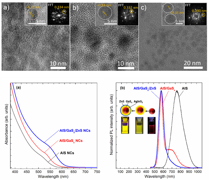
Figure 5. HRTEM images of (a) AgInS₂, (b) AgInS₂/GaSₓ, and (c) AgInS₂/GaSₓ/ZnS; absorption and photoluminescence spectra of AgInS₂, AgInS₂/GaSₓ, and AgInS₂/GaSₓ/ZnS.
We observe a red-shift of the absorption edge toward longer wavelengths after coating the AgInS₂ core with GaSₓ and with GaSₓ/ZnS, corresponding to band-edge energies of ~2.40 eV (517 nm), ~2.33 eV (532 nm), and ~2.26 eV (549 nm), respectively. This red shift may be associated with size increase during shell growth and/or the formation of a core/shell structure in which leakage of the electron–hole wavefunctions into the shell layers reduces quantum confinement.
Uncoated AgInS₂ quantum dots exhibit broad photoluminescence (PL) centered at ~756 nm, with FWHM ~140 nm and a photoluminescence quantum yield (QY) of ~28%. After GaSₓ shelling, the PL becomes very narrow (FWHM ~45 nm), featuring an excitonic peak at ~575 nm along with a small shoulder at lower energy (1.75 eV, 710 nm), and the QY increases to 35%. Remarkably, adding an outer ZnS shell yields purely excitonic emission at 575 nm without the broad low-energy band, and the QY further rises to 60%. Thus, the outermost ZnS shell nearly fully passivates/neutralizes surface dangling bonds, enhancing carrier confinement in AgInS₂ and thereby improving exciton radiative recombination and QY. In addition, the ZnS shell renders AgInS₂/GaSₓ/ZnS more chemically stable, with optical properties essentially unchanged after 12 months under ambient storage. Detailed results are presented in Nanotechnology 33 (2022) 355704 and Nanotechnology 34 (2023) 315601
Noise reduction and feature enhancement in X-ray diffraction data using linear and nonlinear filtering
The maximum-entropy filtering method proposed by Burg in 1967 was designed to extract harmonic frequencies from the noise of stationary time series. Recently, this method has been shown to embody a true maximum-entropy solution in which coefficients within the information band are fully retained, while higher-order coefficients within the white-noise band are replaced by maximum-probability extrapolations from the lower-order coefficients. While such extrapolation mitigates apodization, the refined maximum-entropy approach goes further: it provides model-independent estimates of the locations of singular points that give rise to features in the data.The question addressed here is whether the same advantages—noise suppression, feature detection, and precise feature localization—demonstrated in UV/VIS applications can be realized for X-ray diffraction analysis. The results indicate that they can (Figure 6). Detailed findings are reported in J. Vac. Sci. Technol. B 41 (2023) 044004.
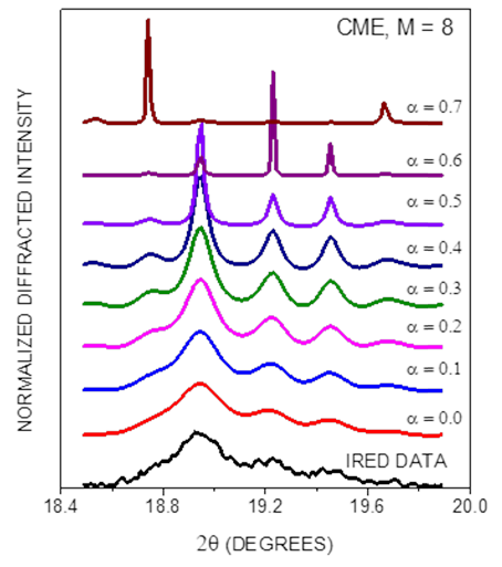
Figure 6. Feature enhancement with increasing α.
SELECTED PUBLICATIONS
1. Tiep Khac Nguyen, Anh D.Kieu, Minh Duc Tran, Thi Thuong Huyen Tran, Cong Doanh Sai, Duc Trong Tran, Diep Ngoc Dang, Son Anh Pham, Huy Hoang Do, Journal of Photochemistry and Photobiology A: Chemistry, "Core-shell building blocks of nanosized Beeswax-Cu2O composites with multifunction of Antibiotic, anti-biofim and self-cleaning", 452 (2024) 115540, IF = 4,1.
2. Thi Thu Hien Bui, Pham Tran Anh Nguyen, Thanh Mai Vu, Thi Huong Giang Tran, Thi Kim Chi Tran and Thi Thuong Huyen Tran*. Mater. Res. Express "Simple precipitation synthesis and solar light-driven photocatalytic degradation of Ag3PO4 floating photocatalysts"11 ( 2024) 095502. IF = 1,8.
3. Cong Doanh Sai, Van Thanh Pham, Thi Ngoc Anh Tran, Thi Thuong Huyen Tran*, Thi Bich Ngoc Vu, Thi Huong Hue Hoang, Anh Son Pham, Thi Minh Thuy Nguyen, Thi Thu Hoai Duong and Huy Hoang Do, Materials Transactions, “Construction of Highly Condensed Cu2O/CuO Composites on Cu Sheet and Its Photocatalytic in Photodegradation of Hazardous Colouring Agent Rose Bengal”, Vol. 64, No. 9 (2023), 2134-2142. IF = 1.377.
4. Thi Thuong Huyen Tran*, Thi Kim Chi Tran, Thi Quynh Xuan Le, Nhat Linh Nguyen, Thi Minh Thuy Nguyen, Thi Thu Hien Pham, Truong Son Nguyen, Hoang Tung Do, Huy Hoang Do. “Engineering the surface structure of brookite‑TiO2 nanocrystals with Au nanoparticles by cold‑plasma technique and its photocatalytic and self‑cleaning property”, J. Nanopart. Res. (2023) 25:203. IF = 2.533.
5. H. H. Do, T. K. C. Tran, T. D. T. Ung, N. T. Dao, D. D. Nguyen, T. H. Trinh, T. D. Hoang, T. L. Le, T. T. H. Tran*, Journal of Water Process Engineering 43 (2021) 102319. Controllable fabrication of photocatalytic TiO2 brookite thin film by 3D-printing approach for dyes decomposition.
6. Nguyen Thu Loan, Tran Thi Thu Huong, Ung Thi Dieu Thuy, Peter Reiss and Nguyen Quang Liem*, Adv. Nat. Sci.: Nanosci. Nanotechnol 15 (2024) 043002. ZnS shelling as the outermost layer on luminescent nanocrystals: a key strategy for quantum dot light-emitting diodes.
7. Ung Thi Dieu Thuy*, Tran Ngoc Huan*, S. Zanna, K. Wilson, Adam F. Lee, N.-D. Le, J. Mensah, V. D. B. C. Dasireddy and Nguyen Quang Liem*, RSC Adv. 14 (2024) 3489-3497. Cu and Zn promoted Al-fumarate metal organic frameworks for electrocatalytic CO2 reduction.
8. N. T. Loan, U. T. D. Thuy, N. Q. Liem* (2023). Fluorescent Biosensors Based on II–VI Quantum Dots. In: Korotcenkov, G. (eds) Handbook of II-VI Semiconductor-Based Sensors and Radiation Detectors. Springer, Cham. https://doi.org/10.1007/978-3-031-24000-3_18.
9. Hoang V Le, Thuy T D Ung*, Phong D Tran, Huy V Mai, Bich D Do and Liem Q Nguyen*, J. Phys. D: Appl. Phys. 56 (2023) 465502. Enhancing photoelectrocatalytic activity and stability of p-Cu2O photocathode through n-TiO2 coating for improved H2 evolution reaction.
10. N. T. Loan, T. T.T. Huong, L. M. Anh, L. V. Long, H. Han, U. T. D. Thuy*, N. Q. Liem*, Nanotechnology 34 (2023) 315601. Double-shelling AgInS2 nanocrystals with GaSx/ZnS to make them emit bright and stable excitonic luminescence.
11. T. T.T. Huong, N.T. Hiep, N. T. Loan, L. V. Long, H. Han, N. T. Thao, U. T. D. Thuy*, N. Q. Liem, Adv. Nat. Sci.: Nanosci. Nanotechnol 14 (2023) 025017. Improved stability and luminescent efficiency of AgInSe2 nanocrystals by shelling with ZnS.
12. N. T. Loan, N. T. Hiep, T. T. T. Huong, U. T. D. Thuy*, T. T. T. Huyen, D. L. H. Tan and N. Q. Liem, Adv. Nat. Sci.: Nanosci. Nanotechnol 13 (2022) 045012. Al-fumarate metal-organic frameworks adsorbent for removal of organic compound and gas storage.
13. T. T.T. Huong, N. T. Loan, L. V. Long, T. D. Phong, U. T. D. Thuy*, N. Q. Liem*, Optical Materials 130 (2022) 112464. Highly luminescent air-stable AgInS2/ZnS core/shell nanocrystals for grow lights.
14. T. T.T. Huong, N. T. Loan, U. T. D. Thuy*, N. T. Tung, H. Han, N. Q. Liem*, Nanotechnology 33 (2022) 355704. Systematic synthesis of different-sized AgInS2/GaSx nanocrystals for emitting the strong and narrow excitonic luminescence.
15. T. T. T. Huong, N. T. Loan, D. X. Loc, U. T. D. Thuy*, O. Stoilova* and N. Q. Liem, Optical Materials 113 (2021) 110858. Enhanced luminescence in electrospun polymer hybrids containing Mn-doped ZnSe/ZnS nanocrystals.
16. Long Van Le*, Tae Jung Kim, Young Dong Kim, D. E. Aspnes, Physica Status Solidi B 260 (2023) 2200271. Detection of the Biexciton of Monolayer WS2 in Ellipsometric Data: A Maximum-Entropy Success.
17. Long Van Le*, Jeroen A. Deijkers, Young D. Kim, Haydn N. G. Wadley, D. E. Aspnes, Journal of Vacuum Science & Technology B 41 (2023) 044004. Noise reduction and peak detection in x-ray diffraction data by linear and nonlinear methods.
MAJOR EQUIPMENT
|
No.
|
Equipment
|
Features and capabilities
|
Location
|
Operator in charge
|
|
1
|
High‑sensitivity spectrometer (Horiba iHR 550)
|
focal length 550 mm; f/6.4; 300–1000 nm; accuracy ±0.2 nm
|
Room 210, A2
|
Ứng Thị Diệu Thúy
Nguyễn Thu Loan
Lê Văn Long
|
|
2
|
STREAK CAMERA (Optronis, Đức)
|
8‑mm photocathode; temporal resolution <2 ps; spatial resolution >40 lp/mm at the photocathode; synchronous sweep 25–250 MHz
|
Room 210, A2
|
Ứng Thị Diệu Thúy
Nguyễn Thu Loan
Lê Văn Long
|
|
3
|
Femtosecond laser system (Coherent)
|
wavelength 680–1040 nm; SHG/THG; pulse width and intensity depend on wavelength
|
Room 210, A2
|
Nguyễn Thu Loan
Ứng Thị Diệu Thúy
Lê Văn Long
|
|
4
|
Spectroscopic ellipsometer (Horiba)
|
190–2100 nm; determines dielectric function (refractive index), thin‑film thickness, and related properties
|
Room 210, A2
|
Lê Văn Long
Nguyễn Thu Loan
Ứng Thị Diệu Thúy
|
|
5
|
Dual‑detector gas chromatograph (Claurus 690, PerkinElmer)
|
FID and TCD; oven 50–450 °C; ramp 0–160 °C/min; split/splitless injector for FID
|
Room 218, A2
|
Ứng Thị Diệu Thúy
Trần Thị Thu Hương
|
|
6
|
Potentiostat SP-300 (Biologic, France)
|
2 channels; ±500 mA; control ±10 V; compliance ±12 V; voltage resolution 1 µV; current resolution 760 fA; 10 µHz–7 Hz; RDE/RRDE, QCM connectivity
|
Room 218, A2
|
Ứng Thị Diệu Thúy
Trần Thị Thu Hương
Nguyễn Minh Thư
|
|
7
|
Potentiostat SP-50 (Biologic, France)
|
1 channel; acquisition time 200 µs (EC‑Lab); output ranges ±2.5/±5/±10 V
|
Room 210, A2
|
Trần Thị Thu Hương
Lê Văn Long
Nguyễn Minh Thư
|
|
8
|
Dilatometer (Linseis)
|
measures ΔL and CTE from room temperature to 1400 °C; ΔL range 100–5000 µm
|
Room 217, A2
|
Lê Văn Long
Trần Thị Thu Hương
Ứng Thị Diệu Thúy
|
|
9
|
Spray pyrolysis system (Holmarc, India)
|
reservoirs 50 and 250 mL; X‑axis 10–800 mm/s; Y‑axis 1–12 mm/s; max travel (X–Y) 200 mm; substrate 150×150 mm; max 500 °C; 230 V, 50 Hz
|
Room 218, A2
|
Trần Thị Thu Hương
Nguyễn Minh Thư Ứng Thị Diệu Thúy
|